Medical Devices
Minimally Invasive Spinal Fusion
WARF: P140303US01
Inventors: Nathaniel Brooks, Meghan Anderson, Hannah Meyer, Hannah Pezzi, Amy Slawson, Andrea Schuster
The Wisconsin Alumni Research Foundation (WARF) is seeking commercial partners interested in developing a less invasive method for delivering bone graft material to the spine of a patient.
Overview
An estimated eight out of 10 people will be affected by a spine condition during their lifetime, making treatment options like spinal fusion surgery imperative. Spinal fusion is a surgical procedure in which vertebrae along the spinal column are fused and immobilized. The procedure can reduce back pain and help treat injuries or deformities.
In general, the procedure involves joining two or more vertebrae together using temporary stabilizing hardware (e.g., screws, rods) and bone graft material that provides additional support as the hardware inevitably loosens over time. Presently, inserting a bone graft is a highly invasive process, requiring pulling back the body tissue and laying the graft in by hand.
A less intrusive technique could ease patients’ pain, minimize scarring, shorten hospital stays and reduce healthcare costs.
The Invention
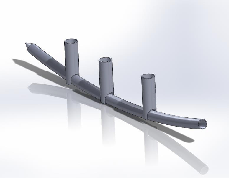
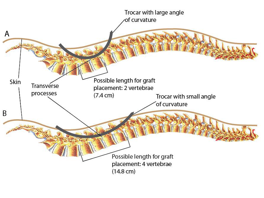
UW–Madison researchers have developed a method and surgical instrumentation to enable minimally invasive spinal fusion. In the procedure, a sharp pointed tube (a trocar) is inserted along the spine. Using the trocar as a guide, the bone graft material is pulled into place. The trocar is removed, releasing the graft in the desired location along the spine.
Applications
- Spinal fusion therapy
- Surgical instrument kit
Key Benefits
- New method is minimally invasive.
- No comparable procedure or device is currently available.
- Faster healing time, reduced costs and less pain for patients
Stage of Development
A prototype has been fabricated and tested in a tissue-mimic phantom. Results demonstrate that the new design allows for successful insertion of the graft within 2 millimeters of the desired location.
Tech Fields
For current licensing status, please contact Jeanine Burmania at [javascript protected email address] or 608-960-9846
Figures