Research Tools
Methods for Bioprinting Functional Human Neural Tissues
WARF: P220006US02
Inventors: Su-Chun Zhang, Yuanwei Yan
The Wisconsin Alumni Research Foundation is seeking commercial partners interested in employing an innovative, customizable method regulating the production of a functional human neural tissue model. Using a standardized platform to produce functional human neural tissue enhances the model’s replicability for targeted neurological disease study and drug screening.
Overview
Su-Chun Zhang, a world-renowned neuroscientist and pioneer in stem cell research, has developed a method for printing neural tissues that allows cells to form neural networks in vitro. The human brain is complex, containing a multitude of specialized neuronal and glial cells that create a neural network responsible for key functions such as cognition, consciousness and reasoning. Studying neuron-glia interactions is critical for investigating neurological diseases including Alzheimer’s disease, Parkinson’s disease and autism spectrum disorders.
However, studying these interactions remains challenging due to the lack of a reliable model representing living human neural tissues. While brain organoids generated from human pluripotent stem cells are a viable solution, functional neural networks in this system are not formed until months to over a year later, and the circuits that are formed are randomly positioned. An alternative solution that complements the organoid system is 3-D bioprinting, enabling the assembly of human tissues by spatially controlling the deposition of living cells and hydrogels to generate biologically complex cytoarchitecture. While printing neural tissues is not novel, this method enables printed tissues to form functional neural networks in a fraction of the expected time.
However, studying these interactions remains challenging due to the lack of a reliable model representing living human neural tissues. While brain organoids generated from human pluripotent stem cells are a viable solution, functional neural networks in this system are not formed until months to over a year later, and the circuits that are formed are randomly positioned. An alternative solution that complements the organoid system is 3-D bioprinting, enabling the assembly of human tissues by spatially controlling the deposition of living cells and hydrogels to generate biologically complex cytoarchitecture. While printing neural tissues is not novel, this method enables printed tissues to form functional neural networks in a fraction of the expected time.
The Invention
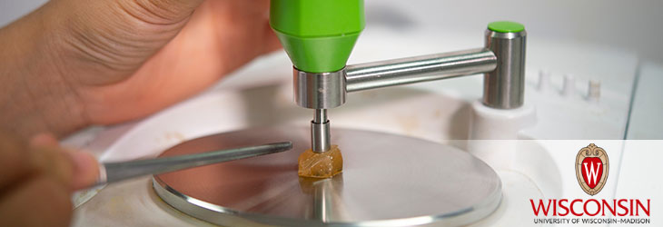
UW-Madison researchers have developed methods for bioprinting a human neural network in vitro. Their method fabricates human neural tissues that form a functional neural network within weeks. They developed a bioink (printing gel) that can be combined with nearly mature cell progenitors of the desired cell types to form a 3-D structure amenable to morphological and functional assessment. The bioink promotes neural cell survival and differentiation so that printed neural tissues mature and form functional networks within a month, and the resulting tissues display expected neural function under physiological and pathological conditions for at least two months after preparation.
The researchers generated neuronal and astrocyte progenitors from human pluripotent stem cells using previous protocols from their and other labs, confirming the cell identity using cell surface markers (e.g., GABA for GABA interneuron progenitors, FOXG1 and PAX6 for cortical progenitors, S100 and GFAP for astrocyte progenitors, etc.). One month after tissue formation, they validated that the neurons exhibited pyramidal morphology and formed dendritic structures. Furthermore, functional assessments confirmed neuronal maturation in the printed tissue, as well as the formation of functional neural networks with functionally integrated astrocytes. The researchers used an inexpensive 3-D bioprinter, but other more complex units would also work.
The researchers generated neuronal and astrocyte progenitors from human pluripotent stem cells using previous protocols from their and other labs, confirming the cell identity using cell surface markers (e.g., GABA for GABA interneuron progenitors, FOXG1 and PAX6 for cortical progenitors, S100 and GFAP for astrocyte progenitors, etc.). One month after tissue formation, they validated that the neurons exhibited pyramidal morphology and formed dendritic structures. Furthermore, functional assessments confirmed neuronal maturation in the printed tissue, as well as the formation of functional neural networks with functionally integrated astrocytes. The researchers used an inexpensive 3-D bioprinter, but other more complex units would also work.
Applications
- Provides a platform that rapidly and reliably generates a functional human neural tissue model for neurological disease study and drug screening
- Standardizes bioprinting of neural tissue such that the anatomical and potentially functional properties of the printed tissues are predictable depending on the composition of the printed progenitor cells
Key Benefits
- No comparable model is currently available on the market
- Offers a unique in vitro method for creating human neural tissue containing a functional neural network
- Provides the shortest available turnaround time to generate a functional neural network in human neural tissues after bioprinting
- Uses customizable bioprinting options that expand research possibilities and increase the health and survivability of printed human neural tissue specimens
Stage of Development
The researchers’ innovative method for constructing human neural tissue containing a functional neural network suggests tangible opportunities for neurological studies and drug testing applications. Their customizable bioprinting options yield a multi-layered structure containing neural progenitor cells that are positive for MAP2, a neural cell marker. Over extended culture, the neurites become denser and form numerous synapses, demonstrating that the printed neural tissue retains a stable structure and has the capacity to form a neural network. Additionally, their bioprinting methods have been used to model Alexander disease (AxD) in printed human neural tissues. As expected, neural tissues printed with AxD astrocytes showed significantly fewer calcium responses than the isogenic control. Their results indicate that the disease-relevant functional phenotypes are detectable in the printed human neural tissues.
Additional Information
For More Information About the Inventors
Tech Fields
For current licensing status, please contact Andy DeTienne at [javascript protected email address] or 608-960-9857